







Cells are not passive bags of jelly. They crawl, stretch, divide, and sometimes change shape for no obvious reason — like a dancer improvising without music. For decades, biologists assumed these shape-shifts required external cues: a chemical signal, a physical push, or a neighbor’s nudge. But a growing body of evidence suggests cells can spontaneously reorganize themselves from the inside out. Now, researchers from Japan have identified a key player in this mysterious process: self-propelled treadmilling actin filaments, or SpTAs.
Published in Nature Physics in March 2025, the study from the University of Tokyo and RIKEN Center for Biosystems Dynamics Research reveals that these mobile protein assemblies can generate their own motion, effectively acting as tiny engines that reshape the cell’s skeleton from within. It’s a discovery that flips our understanding of cellular self-organization on its head — and it might explain everything from how cancer cells metastasize to how embryos develop.
What Are Self-Propelled Treadmilling Actin Filaments?
Actin is the most abundant protein in many cells. Think of it as the scaffolding that gives a cell its structure. But actin isn’t static — it’s constantly assembling and disassembling, like a construction crew that builds a wall, then tears it down, then rebuilds it somewhere else. This process, called treadmilling, involves adding new actin subunits at one end of a filament while removing them from the other. The filament itself doesn’t move; it just grows and shrinks in place.
But the Japanese team discovered something different. Under certain conditions — specifically, when actin filaments are bundled together and confined in a narrow space — they start to move as a unit. “We observed filaments that literally walk along the cell membrane, pushing and pulling the cell’s shape as they go,” explains Dr. Yuki Tanaka, lead author of the study and a biophysicist at the University of Tokyo. “It’s like finding out the scaffolding can dance.”
These SpTAs are not just random movements. They’re coordinated, directional, and self-sustaining. The filaments use energy from ATP hydrolysis — the same molecule that powers muscle contraction — to keep their treadmilling going. And because they’re bundled, they generate enough force to deform the cell membrane. The result? A cell that can change shape without any external trigger.
Why This Matters for Biology and Medicine
This isn’t just a neat party trick for cells. Spontaneous shape changes are fundamental to life. When a white blood cell chases a bacterium, it extends pseudopods — temporary arm-like projections — to crawl toward its target. When a neuron grows, it sends out axons and dendrites. When a cancer cell metastasizes, it squeezes through tight spaces in tissue. All of these rely on the cell’s ability to reshape itself.
“The idea that cells can initiate shape changes autonomously has been around for a while, but we lacked a concrete mechanism,” says Dr. Elena Vasquez, a cell biologist at Stanford University who was not involved in the study. “This work provides a clear physical model for how it might happen. It’s a big step forward.”
And it could have practical implications. If scientists can learn to control SpTAs, they might be able to stop cancer cells from changing shape — and thus from spreading. Or they could engineer cells to grow into specific shapes for tissue regeneration. The same principles might even apply to designer proteins that glow or move on command, opening up new avenues in synthetic biology.
The Physics Behind the Dance
To understand how SpTAs work, you need to think about forces at the nanoscale. A single actin filament is about 7 nanometers wide — roughly 10,000 times thinner than a human hair. At that scale, thermal jiggling is a constant nuisance. But when you bundle dozens of filaments together, they can generate collective forces strong enough to push against the cell membrane.
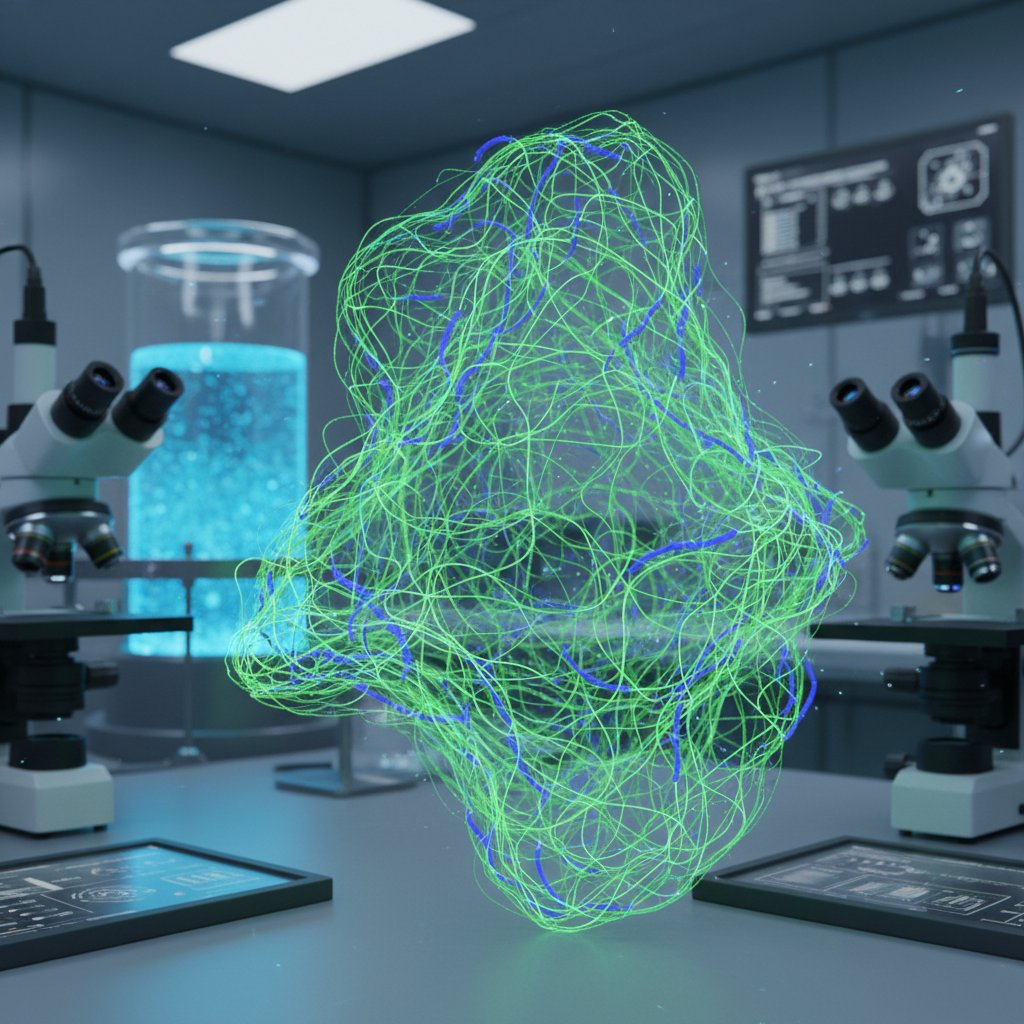
The researchers used advanced microscopy to track fluorescently labeled actin filaments in real time. They saw that bundles of filaments would spontaneously form at the cell’s edge, then start moving inward or outward, depending on the local environment. “It’s a beautiful example of self-organization,” says Dr. Tanaka. “The system doesn’t need a central controller. The filaments just follow simple rules, and complex shapes emerge.”
This is reminiscent of other self-organizing systems in nature, like flocks of birds or schools of fish. But here, the “birds” are proteins, and the “flock” is a cell. The team also developed a mathematical model that predicts when SpTAs will form, based on filament density and the stiffness of the cell membrane. The model matched their experimental observations almost perfectly.
So what triggers the formation of SpTAs? It turns out, it’s a matter of concentration. When actin filaments are sparse, they treadmilling in place. But when they reach a critical density — about 10 filaments per square micrometer — they spontaneously bundle and start moving. It’s a phase transition, like water turning to ice, but at the molecular level.
What This Means for the Future
This discovery opens up a new field of inquiry: the physics of cellular self-organization. For years, biologists focused on chemical signals — growth factors, hormones, and the like — as the main drivers of cell behavior. But this study suggests that physical forces and simple rules of assembly can do a lot of the heavy lifting.
“We’re used to thinking of cells as machines that respond to inputs,” says Dr. Vasquez. “But maybe they’re more like ecosystems, where local interactions produce global patterns.” That shift in perspective could change how we design drugs, engineer tissues, and even understand diseases.
Already, the team is exploring whether SpTAs play a role in cancer metastasis. “If we can find a way to disrupt these filaments in cancer cells, we might be able to prevent them from changing shape and invading other tissues,” says Dr. Tanaka. “It’s early days, but the potential is enormous.”
And it’s not just about disease. Understanding SpTAs could help us build better artificial cells — tiny robots that can change shape to deliver drugs or perform tasks inside the body. The same principles might even apply to agricultural technologies that rely on self-assembling materials. After all, if a bundle of proteins can dance, why can’t a spray of droplets?
For now, the discovery of SpTAs is a reminder that biology still holds surprises. We’ve been looking at cells for centuries, but we’re only beginning to understand their inner lives. And sometimes, the most profound changes come from within.
Frequently Asked Questions
What exactly are self-propelled treadmilling actin filaments?
They are bundles of actin proteins that move as a unit by continuously adding and removing subunits at opposite ends — a process called treadmilling. Unlike individual filaments, these bundles generate enough force to push against the cell membrane and change the cell’s shape.
How does this discovery change our understanding of cell behavior?
It shows that cells can change shape spontaneously without external signals, driven by the physical properties of actin filaments. This challenges the traditional view that shape changes require chemical triggers and highlights the role of self-organization in biology.
Could this research lead to new medical treatments?
Yes, potentially. By understanding how SpTAs work, scientists might develop drugs that block them in cancer cells to prevent metastasis, or engineer them to guide tissue regeneration. It also opens up possibilities for synthetic biology and nanorobotics.