








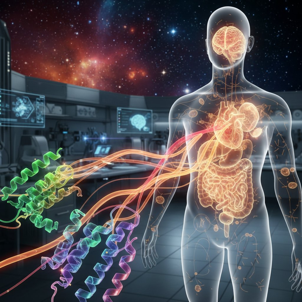
For decades, biologists have relied on fluorescent proteins like GFP to light up cells. But their glow fades beyond a few millimeters of tissue. Now, an international team led by researchers at the National Center for Tumor Diseases (NCT/UCC) in Dresden has engineered proteins that emit light in the near-infrared (NIR) and short-wave infrared (SWIR) spectrum—allowing imaging through centimeters of tissue for the first time. It’s a quantum leap for medical imaging.
Published in Nature Biotechnology, the study introduces a family of designer proteins that are not only brighter than anything nature offers but also genetically encodable. That means scientists can insert the genes for these proteins into specific cells—cancer cells, immune cells, neurons—and watch them glow from outside the body. “We’ve essentially created a molecular flashlight that can shine through an entire organ,” says Dr. Oliver Bruns, a biophysicist at NCT Dresden and senior author of the study.
The Challenge: Seeing Through the Fog of Living Tissue
Biological tissue is murky. Water, lipids, and hemoglobin all absorb and scatter visible light, which is why you can’t see your own liver from the outside. Traditional fluorescent proteins like GFP (green fluorescent protein) work beautifully in a petri dish or a thin slice of tissue, but their signal drops to almost nothing beyond a millimeter or two. Near-infrared light (700–1000 nm) and short-wave infrared (SWIR, 1000–1700 nm) travel much farther because they encounter less scattering and absorption. Synthetic dyes that emit in these ranges have been around for years, but they’re toxic, can’t be made by cells, and fade quickly. “The holy grail has been to create a genetically encoded fluorescent protein that works in the SWIR window,” explains Dr. Bernardo Arús, a researcher at NCT/UCC and co-first author. “Synthetic dyes work, but they can’t be targeted to specific cells without complex chemistry. Our proteins are made by the cells themselves.”
So the team turned to a class of light-sensitive proteins called phytochromes, which naturally absorb NIR light in plants and bacteria. By mutating the protein structure and swapping in different chromophores (the light-absorbing molecule inside), they shifted the emission peak from the red edge into the SWIR region. The result: a protein they call miRFP (monomeric infrared fluorescent protein) that emits at 1100 nm—deep in the SWIR band. In mouse models, the team could image tumors buried 2 centimeters deep, something impossible with any previous fluorescent protein.
Engineering a Brighter, Deeper Glow
But engineering a protein that emits at longer wavelengths is only half the battle. The chromophore—biliverdin, a natural byproduct of heme breakdown—must be supplied in sufficient quantity. Mammalian cells don’t make much of it, so the researchers also co-expressed an enzyme that boosts biliverdin levels. That tweak increased brightness tenfold. “It was like adding a turbocharger to an already powerful engine,” says Bruns. The team then tested their proteins in living mice, injecting tumors with cells expressing miRFP and imaging them with a custom-built SWIR camera. The images were startlingly clear: blood vessels, lymph nodes, and even single cancer cells glowed through layers of skin and muscle.
This isn’t the first time scientists have used SWIR for imaging—synthetic quantum dots and carbon nanotubes have been used for years. But those materials are inorganic and can’t be genetically targeted. “The real breakthrough is that we can now tag any protein of interest with a SWIR-emitting tag,” says Arús. “We can watch how a cancer cell moves, how an immune cell attacks, or how a neuron fires—all in real time, deep inside a living animal.” The team has already used the proteins to track glioblastoma cells in the brains of mice, showing that the tumors invade along blood vessels—a behavior that was previously invisible with standard GFP imaging. For context, similar nanoparticle-based approaches are being explored for human brain cancer, as reported in sugar-coated nanoparticles that target the deadliest brain cancer.
From Bench to Bedside: What This Means for Patients
The immediate applications are in preclinical research—drug development, cancer biology, immunology. But the long-term potential for human medicine is enormous. Imagine a surgeon injecting a patient with a virus that carries the gene for miRFP into tumor cells. During surgery, she dons special goggles that let her see SWIR light, and the tumor glows bright green (or rather, false-colored green) against healthy tissue. No guesswork. No need for radioactive tracers. Or consider a diabetes patient: a small implant of engineered cells that produce miRFP when blood sugar drops too low could be read by a wearable SWIR detector, alerting the patient before symptoms start. “These proteins are fully biocompatible,” notes Bruns. “They’re made from amino acids, not heavy metals. The body already produces the chromophore. We’re not adding anything toxic.”
That’s a critical advantage over existing contrast agents used in MRI or PET scans, which often involve gadolinium or radioactive isotopes. “Optical imaging is cheaper, faster, and safer,” says Arús. “But until now it was limited by depth. We’ve just pushed that limit from millimeters to centimeters.” The team is now working on making the proteins even brighter and shifting the emission further into the SWIR range (beyond 1300 nm), where tissue is even more transparent. They’re also engineering versions that can be activated by light, allowing for super-resolution imaging deep inside tissue.
The Future of Optical Imaging
But there’s a catch: the current proteins require an external light source to excite them, and that light must penetrate the tissue as well. For deep organs like the pancreas or heart, the excitation light may not reach enough to produce a strong signal. The team is tackling this by engineering bioluminescent versions—proteins that produce their own light without an external source, like firefly luciferase but in the SWIR range. “That’s the next frontier,” says Bruns. “If we can make these proteins glow without needing to shine a light on them, we could image anywhere in the body.” The work is still in animal models, but the researchers are already in talks with clinicians to test the proteins in human tissue samples. “We’re probably a few years away from first-in-human trials,” Arús estimates. “But the path is clear.”
For now, the study marks a watershed moment for optical imaging. It’s not every day that a new tool comes along that lets you see the invisible—but these designer proteins do exactly that. And they’re made from the same stuff as life itself.
Frequently Asked Questions
What is SWIR imaging and why is it better than traditional fluorescence?
SWIR (short-wave infrared) imaging uses light between 1000 and 1700 nm. At these wavelengths, biological tissue scatters and absorbs far less light than visible or even near-infrared light. This allows imaging through several centimeters of tissue, whereas standard GFP or visible fluorescence is limited to a few millimeters. SWIR also has lower background autofluorescence, giving much higher contrast.
How are these designer proteins different from synthetic SWIR dyes?
Synthetic SWIR dyes are small molecules that are injected into the body; they cannot be produced by cells, so they cannot be genetically targeted to specific cell types. The new proteins (miRFPs) are genetically encodable—meaning scientists can insert the gene into a cell’s DNA, and the cell will continuously produce the fluorescent protein. This allows long-term tracking of living cells, such as cancer cells or immune cells, without repeated injections.
When will this technology be available for human patients?
The current work is in mouse models. The researchers are now optimizing the proteins for human use, including brightness, stability, and safety. They estimate first-in-human clinical trials could begin within 3–5 years, likely starting with intraoperative imaging during cancer surgery. Regulatory approval and scaling up production will take additional time, but the biocompatibility of the proteins (made from natural amino acids and a common metabolic byproduct) is a major advantage.